Dr. Bhowmik is actively involved in the domain of microscopic cell imaging for early diagnosis of cervical cancer. Cervical Cancer (CC) is the form of malignant disease (i.e. gynaecologic malignancy) that develops in the cells of the cervix or the neck of the uterus. Human Papilloma Virus (HPV) and its numerous strains, around 200 strains, have been identified as a causative factor for cervical cancer [2]. When cervical cancer occurs, cells in the cervix region do not suddenly change into cancer. Instead, the normal cells of the cervix first gradually develop precancerous changes which later turn into cancer. It takes years for this orderly progression to occur and thus cervical cancer is mediciable, if it is detected in early stage and treated appropriately. In clinical practices, conventional pap-smear test continues to be prevalent for prevention and early detection of cervical cancer and have huge inter and intra observer variability. These issues can be addressed by automating the screening methods, which can offer efficient and accurate approaches to analyze vast amount of data and relieve the laborious tasks required by pathologists. To automate the screening methods, extraction of structure information of cells (e.g., shape, staining intensity, texture, and number) is vital to the early detection of cervical cancer. This requires accurate segmentation of nucleus region from the holistic pap-smear images. However, segmentation of suspicious nucleus regions from pap-smear cytological images has remained a challenge to the research community as illustrated in Fig. 1.
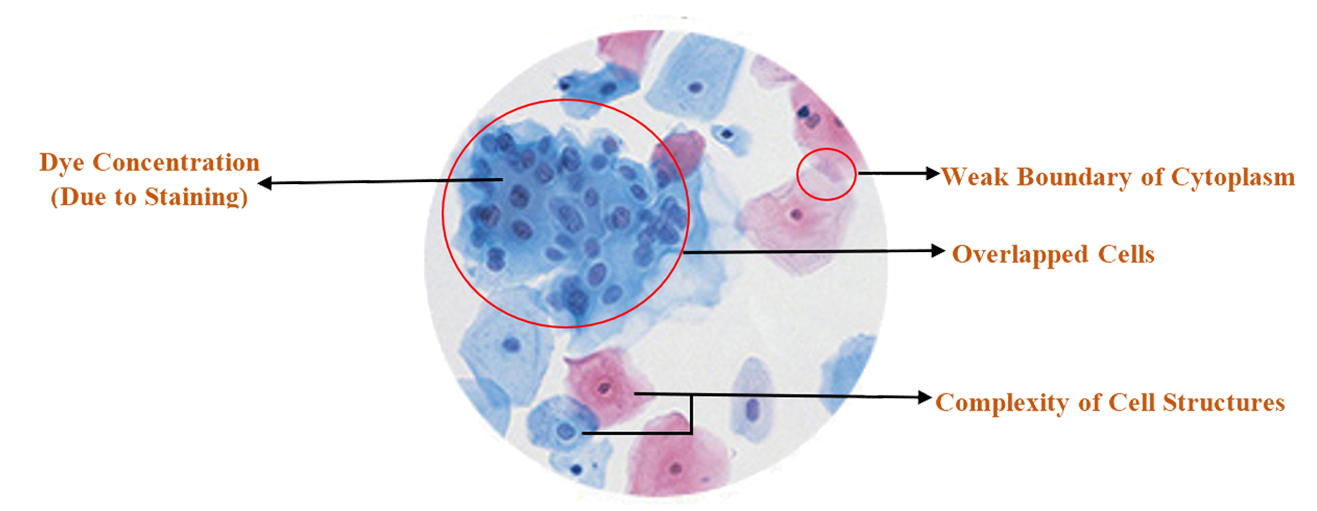
Figure 1. Challenges of Pap-Smear Cytological Images for Cervical Abnormality Detection
- Scope:
In North-East India, due to the inadequate medical facilities,
lack of awareness, lack of expert oncologists and late detection,
the cancer incidence rate doubles the national average,
for which North-East (NE) India is now known as the “Cancer Capital”.
Among all cancers in NE, Cervical Cancer (CC) is the second most common
in female and according to a report on Cancer Burden in North Eastern States of India,
NE shows the CC incidence rate which is more than half of the CC incidence rate in rest of India.
Fixing the north eastern cancer crisis requires cumulative efforts of hospitals,
administrative bodies, diagnostic centers, NGOs to impart the right awareness and
lifestyle guidance. In pursuit of this, our motive is to understand the epidemiology
of CC in NE Region especially in Tripura to control the high incidence and mortality rate
of CC by automatically detecting and treating the disease in early stages.
- Overview of our Research in Early Cervical Cancer Screening :
- AGMC-TU Pap Smear Cytological Image Dataset:
For designing a ground truth annotated microscopic cell image dataset, a standard cell preparation protocol is designed under the supervision of medical expert. By maintaining the standard protocol suite, the pap-smear images (single cell images) were taken randomly from asymptotic population (normal and abnormal subjects) of Tripura. Besides, the ground truth images of suspicious nucleus region in the form of binary masks are also annotated and provided with the dataset. Some sample pap-smear images along with the ground truth images of the suspicious nucleus regions are shown in Figure 2. The key features of this dataset are:
- The dataset contains newly created standardized pap-smear images focusing on cervical cancer screening. Creation of such a dataset will expand the area of research in automated disease diagnosis.
The dataset contains ground truth images of suspicious nucleus region in the form of binary masks. The ground truth marking is performed under the supervision of medical expert.
- The pap-smear images of normal and abnormal subjects are present in the dataset.
- Each pap-smear image of the dataset is labelled by consulting with medical expert in this domain.
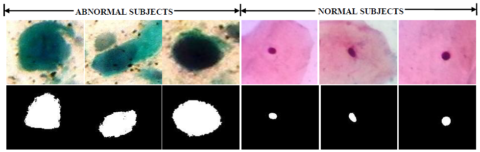
Figure 2. Sample Images of AGMC-TU Pap-Smear Cytological Image Dataset (The top panel represents dataset images and the bottom panel represents corresponding ground truth of dataset images)
- Analysis of AGMC-TU Pap Smear Cytological Image Dataset:
To evaluate the potentiality of microscopic pap-smear cell images in cervical cancer detection, some of our research works including: segmentation of suspicious nucleus regions; feature extraction for asymmetry analysis and classification of normal and abnormal pap-smear images have been done.
- Featured Article(s) in the Proposed Domain:
-
Sourav Dey Roy, Mrinal Kanti Bhowmik, Niharika Nath and Abhijit Datta, "Nucleus Region Segmentation Towards Cervical Cancer Screening Using AGMC-TU Pap-Smear Dataset", Proceedings of 2018 the International Conference on Pattern Recognition and Artificial Intelligence (PRAI 2018), Published by ACM, pp. 44-53, 2018.
-
Mrinal Kanti Bhowmik, Niharika Nath, Abhijit Dutta and Anjan Kumar Ghosh, "Shape Feature Based Automatic Abnormality Detection of Cervico-Vaginal Pap Smears", Journal of Image and Graphics, Published by Engineering and Technology (ETP), Vol. 5, No. 2, pp. 52-58, 2017.
-
Payel Rudra pal, Mrinal Kanti Bhowmik, Debotosh Bhattacharjee, "Automated Cervical Cancer Using PAP SMEAR Images", Proceedings of 4th International Conference on Soft computing for Problem Solving (‘SOCPROS’ 2014), Published by Springer, Vol. 1, pp. 267-278, 2014.